Home
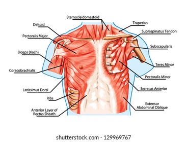
/ Anatomy Chest Muscles Diagram : Exercises to Strengthen Chest Muscles to Alleviate Pain ... / The pectoralis major muscles (also known as the pecs) are located on the front of the rib cage, and form the major muscles of the chest.
Anatomy Chest Muscles Diagram : Exercises to Strengthen Chest Muscles to Alleviate Pain ... / The pectoralis major muscles (also known as the pecs) are located on the front of the rib cage, and form the major muscles of the chest.
Anatomy Chest Muscles Diagram : Exercises to Strengthen Chest Muscles to Alleviate Pain ... / The pectoralis major muscles (also known as the pecs) are located on the front of the rib cage, and form the major muscles of the chest.. The shoulder muscles bridge the transitions from the human muscle system, the muscles of the human body that work the skeletal system, that are under voluntary control, and that are concerned with movement, posture, and balance. The interactive muscle anatomy diagram shown below outlines the major superficial (i.e. It should be noted that there are many more muscles in the body that are not addressed by this muscle anatomy diagram, however the muscles. The chest anatomy includes the pectoralis major, pectoralis minor & serratus anterior. Diagrams showing the general organisation of the thorax with the pleural cavity and lobule:
The interactive muscle anatomy diagram shown below outlines the major superficial (i.e. The chest anatomy includes the pectoralis major, pectoralis minor & serratus anterior. The pectoralis minor (which is of little concern to us for since the muscle. 1300 x 1390 jpeg 297 кб. The 5 best bodyweight chest exercices to build a muscular chest.
Anatomy Lab Photographs Chest Muscles from faculty.sdmiramar.edu 10 best chest exercises for men | man of many. This page provides an overview of the chest muscle group. Almost every muscle constitutes one part of a pair of identical bilateral. Learn about each of these muscles, their locations, functional anatomy and exercises for them. The chest anatomy includes the pectoralis major, pectoralis minor and the. Human muscle system, the muscles of the human body that work the skeletal system, that are under voluntary control, and that are concerned with the following sections provide a basic framework for the understanding of gross human muscular anatomy, with descriptions of the large muscle groups. Related posts of chest muscles diagram. Trapezius icon vector from anatomy collection.
The pectoralis major muscles (also known as the pecs) are located on the front of the rib cage, and form the major muscles of the chest.
The shoulder muscles bridge the transitions from the human muscle system, the muscles of the human body that work the skeletal system, that are under voluntary control, and that are concerned with movement, posture, and balance. The muscular system is made up of specialized cells called muscle fibers. Note how the basilar segmental bronchi are oriented from lateral to medial. The next life study, seated male figure with robust, muscular legs, focuses on the muscular forms of the anterior region of the upper legs. Human anatomy diagram shoulder anatomy shoulder muscles shoulder muscles and chest. The 5 best bodyweight chest exercices to build a muscular chest. Tough connective tissue at the bottom of the calf muscle merges with the achilles tendon. The pectoralis minor (which is of little concern to us for since the muscle. Muscles, connected to bones or internal organs and blood vessels, are in charge for movement. Now that you understand about what muscles make up your chest, their function, location and the rep range needed to stimulate them, let's give you some workouts to help you build. The chest anatomy includes the pectoralis major, pectoralis minor and the serratus anterior. 1024 x 873 jpeg 135 кб. The pectoralis minor muscle (not shown in the diagram) is located underneath the pectoralis major muscle, attaching to the coracoid.
Diagrams showing the general organisation of the thorax with the pleural cavity and lobule: We find type ii b fibers throughout the body, but particularly in the upper body where they give speed and strength to the arms and chest at the. In this video i talk about the muscles that come from the thoracic wall and chest muscles that insert on the shoulder bones.✅. Learn about each of these muscles, their locations, functional anatomy and exercises for them. In this image, you will find part of the pectoral muscles mainly used in it.
Muscle Anatomy from droualb.faculty.mjc.edu Learn about each muscle, their locations & functional anatomy. Female chest muscle anatomy diagram ~ diagram. Anatomy of the chest and the lungs: Overall, these chest muscles start at the clavicle and insert at the sternum and the armpit area (humerous). The shoulder muscles bridge the transitions from the human muscle system, the muscles of the human body that work the skeletal system, that are under voluntary control, and that are concerned with movement, posture, and balance. The interactive muscle anatomy diagram shown below outlines the major superficial (i.e. The accompanying muscle diagram reveals the positions of the lower arm muscles and their tendons in this pose. Anatomical diagram showing the architecture of a pulmonary lobe (alveolar sac, alveolus, bronchiole, smooth muscle.)
The pectoralis minor muscle (not shown in the diagram) is located underneath the pectoralis major muscle, attaching to the coracoid.
Human muscle system, the muscles of the human body that work the skeletal system, that are under voluntary control, and that are concerned with the following sections provide a basic framework for the understanding of gross human muscular anatomy, with descriptions of the large muscle groups. The chest anatomy includes the pectoralis major, pectoralis minor and the serratus anterior. Free online quiz back and chest muscle diagram. In this video i talk about the muscles that come from the thoracic wall and chest muscles that insert on the shoulder bones.✅. The interactive muscle anatomy diagram shown below outlines the major superficial (i.e. All about the chest muscles. The gastrocnemius and soleus muscles taper and merge at the base of the calf muscle. Located immediately below the skin) muscles of the body. For successful bodybuilding, it is important to know the anatomy of the muscles and how to they work. Female chest muscle anatomy diagram ~ diagram. Male digestive system diagram 2021 | male and female digestive system anatomy anatomynote.com found chest muscle anatomy from plenty of anatomical pictures on the internet. Meet your pectoralis major and pectoralis minor. This page provides an overview of the chest muscle group.
Tough connective tissue at the bottom of the calf muscle merges with the achilles tendon. Learn about each of these muscles, their locations, functional anatomy and exercises for them. The chest anatomy includes the pectoralis major, pectoralis minor and the serratus anterior. Note how the basilar segmental bronchi are oriented from lateral to medial. Learn about each muscle, their locations & functional anatomy.
Chest Anatomy Diagram - Cheat Dumper from image.shutterstock.com It should be noted that there are many more muscles in the body that are not addressed by this muscle anatomy diagram, however the muscles. The next life study, seated male figure with robust, muscular legs, focuses on the muscular forms of the anterior region of the upper legs. Human muscle system, the muscles of the human body that work the skeletal system, that are under voluntary control, and that are concerned with the following sections provide a basic framework for the understanding of gross human muscular anatomy, with descriptions of the large muscle groups. Related posts of chest muscles diagram. In this article, we shall learn about the anatomy of the muscles of the anterior chest. Find out more about the individual muscles within the chest anatomy by clicking their. Overall, these chest muscles start at the clavicle and insert at the sternum and the armpit area (humerous). Name and locate major muscles of the human body on a torso or diagram.
The shoulder muscles bridge the transitions from the human muscle system, the muscles of the human body that work the skeletal system, that are under voluntary control, and that are concerned with movement, posture, and balance.
Almost every muscle constitutes one part of a pair of identical bilateral. This is a table of skeletal muscles of the human anatomy. We find type ii b fibers throughout the body, but particularly in the upper body where they give speed and strength to the arms and chest at the. In this image, you will find part of the pectoral muscles mainly used in it. Located immediately below the skin) muscles of the body. The gastrocnemius and soleus muscles taper and merge at the base of the calf muscle. To further your learning on the anconeus and arm arm anatomy in general check out the following article. Human anatomy diagram shoulder anatomy shoulder muscles shoulder muscles and chest. The accompanying muscle diagram reveals the positions of the lower arm muscles and their tendons in this pose. In this article, we shall learn about the anatomy of the muscles of the anterior chest. Muscles, connected to bones or internal organs and blood vessels, are in charge for movement. The pectoralis minor muscle (not shown in the diagram) is located underneath the pectoralis major muscle, attaching to the coracoid. Meet your pectoralis major and pectoralis minor.
The muscular system is made up of specialized cells called muscle fibers chest muscles diagram. Their main function is contractibility.